


单纯疱疹病毒性脑炎(HSE)是一种具有破坏性的中枢神经系统感染性疾病,又称急性坏死性脑炎,是最常见的散发性、致命性脑炎,HSE的病死率高达30%,且神经系统后遗症发生率高[1]。及时启动静脉注射阿昔洛韦进行抗病毒治疗是降低病死率、避免出现严重的神经系统后遗症、改善预后的主要决定因素。目前HSE尚无特效治疗方法,以采用综合治疗,经验性的抗病毒、保护脑细胞及降低颅内压等对症支持治疗为主。CT灌注成像(CTP)可快速显示脑部组织灌注异常及脑组织受损的情况,一般被用于脑卒中的诊断,较少被用于辅助诊断脑炎。本文报道1例CTP辅助诊断单纯疱疹病毒1型 (HSV-1)脑炎的病例以供临床参考。
病例资料
一、主诉、病史及体格检查
患者男,59岁,急性起病,于2022年4月22日因右侧肢体抽搐伴发热半日入本院急诊。患者于入院前半日被他人发现右侧肢体抽搐,呈发作性,每次发作约10 min,发作多次(具体次数不详),伴发热,最高体温为38.5℃,伴精神行为异常,主要表现为躁动不安、骂人、打人及吐口水。考虑患者病情危重,将其由急诊转入重症病房。家属述患者发病前几日有“上呼吸道感染”表现,其他情况无特殊。个人史、家族史、遗传史等均无特殊。入院体格检查:体温38.0℃、脉搏78次/分、呼吸26次/分、血压125/67 mmHg(1 mmHg=0.133 kPa)。患者呈昏睡状态,右侧瞳孔直径3.0 mm、左侧3.0 mm,双侧瞳孔直接和间接对光反射均灵敏,眼球活动正常。四肢肌力查体不合作,肌张力正常,双侧病理征(-),脑膜刺激征(±):颈项强直(±)、Kernig征(-)、Brudzinski征(-)、直腿抬高试验(-)。改良Rankin量表(mRS)评分5分。
二、实验室及辅助检查
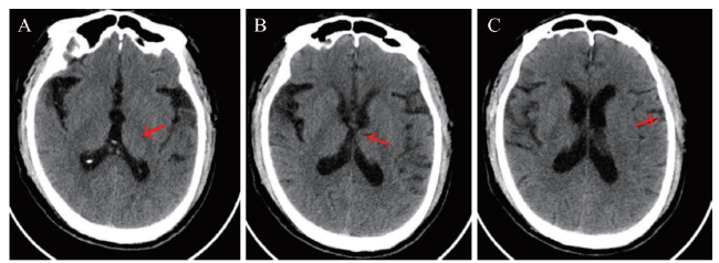
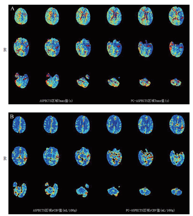
在患者入院24 h内完善相关检查。血常规:血红蛋白 155 g/L、红细胞 4.75×10¹²/L、白细胞 12.51×10⁹/L、中性粒细胞0.78、血小板 189.00× 10⁹/L。降钙素原、肝肾功能、输血前3项(获得性免疫缺陷综合征、丙型肝炎和梅毒)、抗血细胞抗体、抗核提取物抗体、免疫系列等均未见异常。脑脊液检查:腰椎穿刺测得压力190 mmH2O(1 mmH2O = 0.0098 kPa)。脑脊液:白细胞23.00×10⁶/L,单个核细胞百分比 88.70%,多核细胞百分比11.30%;氯117.6 mmol/L,IgG 50.33 mg/L,总蛋白582.15 mg/L;脑脊液病原学检查未见明显异常。患者躁动不安,不能配合完成头颅MRI,遂立即实施头颅CT,可见左侧颞叶皮质肿胀(图1),头颅CT血管成像(CTA)未见颈部血管狭窄,头颅CTP可见左侧颞叶灌注较右侧增加(图2),排除急性脑梗死,不排除感染相关性疾病,初步考虑病毒性脑炎的可能。肥达反应+外斐反应、新型隐球菌荚膜多糖抗原检测(GXM,血清)均为(-)。动态脑电图:背景活动以7~8 Hz慢a波为主,右额颞区可见间隔0.8~2.0 s三相波、尖慢复合波呈周期性发放。重度异常脑电图(爆发-抑制)。脑脊液病原体二代测序(NGS)结果于入院后第3日回报,明确诊断为HSV-1感染所致的HSE。
三、治疗方法及临床转归
根据CTP的提示,于入院后12 h内给予患者阿昔洛韦(0.5 g、每8 h泵注1次)抗病毒治疗、甘露醇降颅压、丙戊酸钠抗癫痫、头孢唑肟抗感染、丙泊酚镇静,并予吸氧及其他对症治疗。入院第3日后患者精神好转,发热高峰下降,但仍偶有踢床和挣扎动作。入院第3日NGS结果回报并确诊HSE后,继续予阿昔洛韦抗病毒治疗,予卡马西平、吡仑帕奈控制肢体抽搐,予奥氮平镇静,予头孢唑肟抗感染,予祛痰、护胃对症治疗并加强气道护理以预防并发症的发生。入院第7日患者体温恢复正常,意识障碍改善,精神行为异常明显好转,无癫痫发作,偶有烦躁不安,可部分回答他人的提问。入院第15日患者体温恢复正常,无癫痫发作,仍偶有烦躁不安,复查头颅CT及MRI未见明显异常病灶,出院mRS评分1分。出院后1个月随访情况良好。
讨论
HSE是由疱疹病毒引起的以精神和意识障碍为主要表现的中枢神经系统感染性疾病,是病毒对脑实质的损伤,其主要病理改变为脑组织的肿胀、坏死、出血、炎细胞浸润,常见于双侧额顶叶、基底节区,甚至可累及小脑,病灶可对称或不对称分布。HSE病死率高达40%~70% ,HSV是一种嗜神经DNA病毒,包括HSV-1和HSV-2两种血清型,约90%的人类HSE由HSV-1引起[1]。Lee等[2]的研究表明,HSV-1感染患者最常见的临床诊断是HSE,而且由HSV-1引发的HSE患者ICU的入住率高达72.7%,出院时严重神经系统后遗症的发生率为27.3%。由HSV-1引发的HSE患者病情发展迅速,病死率较高,所以及时的诊断与治疗是改善患者预后的关键[3]。
病毒性脑炎的主要确诊试验是脑脊液病毒核酸检测[4]。PCR及一代测序虽能准确地检测出病毒病原体,但是受引物每次所能结合的核酸片段有限的限制,检测所用时间较长,对重症患者来说实用性不高,故不能广泛应用于临床[5]。随着影像学技术的发展,设备质量与性能的提高,影像学检查可为病毒性脑炎的诊断提供更多可靠信息,有助于不能进行免疫学或病原学检查的医疗单位作出正确诊断。有研究者提出MRI是早期辅助诊断脑炎的较为理想的方法,其对HSE早期轻度脑水肿较CT敏感,且易发现颞叶底部的低密度灶[6-7]。MRI在检测HSE异常方面的敏感度为80%~90%,MRI中的扩散加权图像和液体衰减反转回复(FAIR)序列是最敏感的[8⇓-10]。较多研究者强调早期应用MRI以辅助诊断重症病毒性脑炎,从而尽早给予患者有效的抗病毒治疗,提高治愈率。但MRI需要患者高度配合,而且成像时间相对较长,影响成像效果因素较多,因此对于急危重症患者不宜实施MRI,对于有体内植入金属物患者亦不宜实施。本例患者由于存在精神行为异常而未能完善头颅MRI,但头颅CTP提供了诊断依据,没有因此耽误患者的治疗。CTP能否替代MRI成为重症病毒性脑炎患者早期启动抗病毒治疗的有力影像学证据值得探讨。对于颅脑病变,多层螺旋CT(MSCT)可以快速且精确显示患者颅内结构的变化,为确诊提供相关影像学证据[11]。MSCT具有快速、方便、检查费用低等优点,能清晰显示颅脑不同位置的横断面解剖关系以及组织结构[12]。有研究者发现MSCT对病毒性脑膜炎诊断准确率可达96.67%[13-14]。岳甜甜[15]认为MSCT诊断病毒性脑膜炎阳性率较高原因可能是其解剖分辨率较高。但也有研究显示,MSCT对重症病毒性脑炎患者的诊断价值并没有高于MRI[16]。CTP在临床中使用较广,可准确地测量大脑、肝脏、胰腺、肾脏等实质脏器。有研究者发现在急性胰腺炎患者中,早期CTP参数显示胰腺短时间的高灌注,随着病情的逐渐进展则表现为相等或者稍低的灌注情况,CTP在中枢神经系统炎症中也显示同样效果[17-18]。CTP对实质脏器细微灌注情况的有效评估是患者病情与预后判断的重要参考。CTP通常用于脑卒中的治疗及预后评估,CTP获取的脑血流量(CBF)、脑血容量(CBV)、对比剂平均通过时间(MTT)和峰值时间(TTP)等血流动力学参数的差异可快速反映脑组织灌注损伤的情况, 且CTP血流动力学参数可有效反映患者血管受累及治疗后再通的情况,可为其预后的评估提供依据-20]。本例的诊断过程提示,CTP对于病毒性脑炎也有重要的诊断价值,病毒性脑炎患者早期CTP可表现为短时间的高灌注,随着病情的逐渐进展可表现为相等或者稍低的灌注情况,CTP既能为HSE提供有力证据,以提高临床早期诊断及治愈率,又能评估预后。但CTP用于脑炎辅助诊断的文献报道较少,其实用性还需要更多的临床研究来证实。
综上所述,通过CTP对患者的脑组织灌注情况进行分析可能有助于病毒性脑炎的诊断,提高确诊率、降低误诊率,从而降低病毒性脑炎患者病死率、改善其预后。