Original Research

Effect of PPAR-γ on proliferation of human pancreatic cancer BxPc-3 cells

  • Li Bo ,
  • Shi Jinglong ,
  • Qiu Houkuang ,
  • Xu Ming
Expand
  • Department of Gastroenterology, Guangdong Second Provincial General Hospital,Guangzhou 510317, China
Xu Ming, E-mail:

Received date: 2021-01-28

  Online published: 2021-10-28

Copyright

Copyright reserved © 2021

Abstract

Objective To evaluate the effect of promoting the expression of peroxisome proliferator-activated receptor-γ (PPAR-γ) on the proliferation of human pancreatic cancer BxPc-3 cells. Methods BxPc-3 cells were cultured and pretreated with PPAR-γ agonist rosiglitazone with a final concentration of 5, 10, 20 and 40 μmol/L, respectively. Western blot was used to detect the expression of PPAR-γ protein. MMT assay was employed to observe the cell proliferation. Results There was no statistical significance in the PPAR-γ expression levels between BxPc-3 cells treated with 5 and 10 μmol/L rosiglitazone and control cells (both P > 0.05). The expression levels of PPAR-γ were gradually up-regulated after treatment with 20 and 40 μmol/L rosiglitazone, which had statistical significance compared with that in the control group (both P < 0.05). The expression level of PPAR-γ in the 40 μmol/L rosiglitazone group was significantly higher than that in the 20 μmol/L rosiglitazone group (P < 0.05). The proliferative activity of BxPc-3 cells treated with 5 and 10 μmol/L rosiglitazone was 0.60±0.07 and 0.57±0.06, which had no statistical significance compared with that in the control group (both P > 0.05). The proliferative activity of BxPc-3 cells treated with 40 μmol/L rosiglitazone was 0.30±0.02, significantly lower than those in the 20 μmol/L rosiglitazone group (0.45±0.03), control group, 5 and 10 μmol/L rosiglitazone groups (all P < 0.05). Conclusion Promoting PPAR-γ expression can effectively inhibit the proliferation of human pancreatic cancer BXPC-3 cells.

Cite this article

Li Bo , Shi Jinglong , Qiu Houkuang , Xu Ming . Effect of PPAR-γ on proliferation of human pancreatic cancer BxPc-3 cells[J]. JOURNAL OF NEW MEDICINE, 2021 , 52(9) : 677 -679 . DOI: 10.3969/j.issn.0253-9802.2021.09.007

胰腺癌是消化系统恶性程度高、预后差的恶性肿瘤之一,由于其侵袭性强,早期往往已经浸润大血管、神经等,造成手术切除难度大,严重降低生存率,5年总体生存率不足1%[1]。过氧化物酶体增殖物-γ(PPAR-γ)除参与脂肪代谢、炎症反应和细胞周期的调控等细胞功能调节外,还与肿瘤发生关系密切[2]。笔者前期试验表明促进PPAR-γ表达可以有效抑制人胰腺癌BxPc-3细胞上皮间充质转化(EMT)及侵袭性[3]。本实验拟明确调控PPAR-γ表达对BxPc-3细胞增殖活性的影响。

材料与方法

一、材料

人胰腺癌BxPc-3细胞购自上海中科院细胞研究所;PPAR-γ激动剂罗格列酮购自美国Sigma;胰酶、高糖DMEM培养基购自美国Gibco 公司;四甲基偶氮唑蓝(MTT)试剂盒购自中国碧云天公司;鼠抗人PPAR-γ抗体及GAPDH内参购自南京凯基公司。

二、方法

1. 细胞株与培养
人胰腺癌BxPc-3细胞用含10%胎牛血清的DMEM培养液于37℃、5%CO2培养箱中培养,细胞均呈贴壁生长。每2 ~ 3 d用0.25%胰酶消化传代1次。根据前期研究经验,将BxPc-3细胞分5组处理96 h,分别为不加药物的对照组(对照组)、加入终浓度5 μmol/L 罗格列酮组(5 μmol/L罗格列酮组)、加入终浓度10 μmol/L 罗格列酮组(10 μmol/L罗格列酮组)、加入终浓度20 μmol/L罗格列酮组(20 μmol/L罗格列酮组)、加入终浓度40 μmol/L 罗格列酮组(40 μmol/L罗格列酮组)。
2. PPAR-γ表达水平的检测
采用蛋白免疫印迹法:收集对数生长期BxPc-3细胞,各组BxPc-3细胞加药后加入600 μl RIPA细胞裂解液,离心30 min,收集上清液即总蛋白,加2倍体积十二烷基酸钠上样缓冲液,电泳后转移至醋酸纤维膜上,脱脂奶粉封闭,加入相应一抗(PPAR-γ抗体,1∶100),4 ℃孵育过夜,洗膜3次,加入羊抗鼠二抗(1∶10 000),37 ℃孵育2 h, 电化学发光(ECL)法检测,感光显影。AlphaImager 2200图像系统分析灰度值,以GAPDH为内参,计算各组细胞的PPAR-γ表达水平。实验组每组设4个平行孔,实验独立重复3次,结果取平均值。
3. BxPc-3细胞增殖活性的检测
采用MTT法检测:收集对数生长期的BxPc-3细胞,各组BxPc-3细胞加药处理后加入20 μl MTT 溶液,孵育4 h,再加入150 μl二甲基亚砜(DMSO)震荡10 min,使用ELISA检测仪于490 nm 波长处测量各组吸光度值(增殖活性)。每组设4个平行孔,实验独立重复3次,结果取平均值。

三、统计学处理

使用SPSS 16.0处理数据。实验数据用 $\bar{x} \pm s$表示,多组间比较采用单因素方差分析,进一步两两比较行LSD-t检验。P < 0.05为差异有统计学意义。

结果

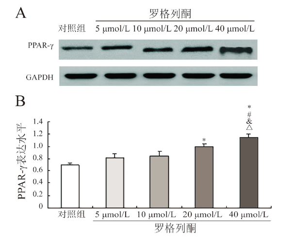
一、不同浓度罗格列酮对BxPc-3细胞PPAR-γ表达水平的影响

5组BxPc-3细胞PPAR-γ表达水平比较差异有统计学意义(F = 40.922,P < 0.001)。5 μmol/L罗格列酮组、10 μmol/L罗格列酮组BxPc-3细胞的PPAR-γ表达水平与对照组比较差异均无统计学意义(P均> 0.05),但20 μmol/L罗格列酮组、40 μmol/L罗格列酮组BxPc-3细胞的PPAR-γ表达水平升高,与对照组比较差异均有统计学意义(P均< 0.05),且40 μmol/L罗格列酮组BxPc-3细胞的PPAR-γ表达水平高于5 μmol/L罗格列酮组、10 μmol/L罗格列酮组和20 μmol/L罗格列酮组(P 均< 0.05),见图1
图1 不同浓度罗格列酮对BxPc-3细胞PPAR-γ表达的影响

A:蛋白免疫印迹法检测结果;B:定量分析结果;与对照组比较*P < 0.05,与5 μmol/L罗格列酮组比较#P < 0.05,与10 μmol/L罗格列酮组&P < 0.05,与20 μmol/L罗格列酮组比较△P < 0.05;n = 4

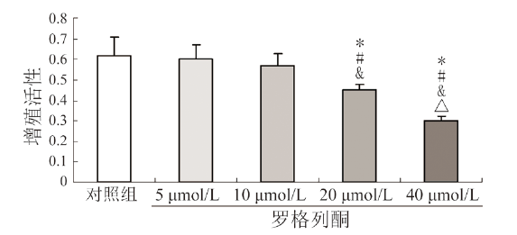
二、不同浓度罗格列酮对BxPc-3细胞增殖活性的影响

MTT检测显示,5组BxPc-3细胞增殖活性比较差异有统计学意义(F = 19.966,P < 0.001)。5 μmol/L罗格列酮组、10 μmol/L罗格列酮处理后BxPc-3细胞增殖活性分别为0.60±0.07、0.57±0.06,与对照组的0.62±0.09比较差异均无统计学意义(P均> 0.05);40 μmol/L罗格列酮组细胞增殖活性为0.30±0.02,低于20 μmol/L罗格列酮组的0.45±0.03,且2组BxPc-3细胞增殖活性均低于对照组、5 μmol/L罗格列酮组和10 μmol/L罗格列酮组(P均< 0.05),见图2
图2 不同浓度罗格列酮对BxPc-3细胞增殖活性的影响

与对照组比较*P < 0.05,与5 μmol/L罗格列酮组比较#P < 0.05,与10 μmol/L罗格列酮组比较&P < 0.05,与20 μmol/L罗格列酮组比较△P < 0.05;n = 4

讨论

PPAR是一类由配体激活的转录因子,有α、β和γ这3种亚型,3种亚型在功能和结构有一定差异[4]。PPAR-γ 配体分为外源性和内源性2大类,其中外源性配体以胰岛素增敏剂噻唑烷二酮类药物为代表,包括罗格列酮、吡格列酮等,临床用于治疗胰岛素抵抗、糖尿病等,内源性配体则包括一些不饱和脂肪酸及其代谢产物,如花生四烯酸、亚油酸等,还有前列腺素衍生物、前列腺素D2等,这些天然激动剂活性都较低[5,6]
PPAR-γ 最初发现是与胰岛素抵抗、脂肪细胞分化和器官纤维化等有关。近年研究显示其与肿瘤发生关系密切。已有报道PPAR-γ在胃癌、结肠癌、胰腺癌、肝癌、肾癌、乳腺癌、食管癌和淋巴瘤等多种恶性肿瘤中表达[7]。Tsujie等(2003年)报道PPAR-γ在包括BxPc-3细胞在内的6种胰腺癌细胞株上表达。Kristiansen等(2006年)在129例胰腺癌患者的癌组织中检测PPAR-γ,71.3%的病例中有PPAR-γ的表达,且其表达与胰腺癌的分级和分期呈正相关,推测PPAR-γ与胰腺癌有关。PPAR-γ在胰腺癌中的确切作用机制尚未阐明。Eibl等(2001年)认为,PPAR-γ可能通过诱导胰腺癌细胞凋亡,进而阻止胰腺癌细胞生长。
笔者前期研究使用罗格列酮促进BxPc-3细胞中PPAR-γ表达,结果显示罗格列酮可以有效抑制人胰腺癌BxPc-3细胞EMT及侵袭性。本实验中笔者继续将不同浓度罗格列酮作用于BxPc-3细胞,结果显示20、40 μmol/L罗格列酮可以有效促进PPAR-γ的表达,且40 μmol/L罗格列酮组BxPc-3细胞的PPAR-γ表达高于20 μmol/L罗格列酮组。MTT结果提示20 μmol/L罗格列酮组、40 μmol/L罗格列酮组BxPc-3细胞增殖活性明显下降,表明PPAR-γ表达可以有效抑制人胰腺癌BxPc-3细胞增殖活性。
综上所述,本研究结果初步揭示了PPAR-γ激动剂的抗胰腺癌细胞增殖特性,而探索PPAR-γ参与人胰腺癌BxPc-3细胞增殖及EMT的信号传导通路将是我们下一步的研究方向。
[1]
Zöller M. Pancreatic cancer diagnosis by free and exosomal miRNA. World J Gastrointest Pathophysiol, 2013, 4(4):74-90.

DOI

[2]
Sabatino L, Pancione M, Votino C, Colangelo T, Lupo A, Novellino E, Lavecchia A, Colantuoni V. Emerging role of the β-catenin-PPARγ axis in the pathogenesis of colorectal cancer. World J Gastroenterol, 2014, 20(23):7137-7151.

DOI

[3]
李博, 施景龙, 魏海雄, 邱厚匡, 许鸣. 促进PPAR-γ表达对TGF-β1诱导人胰腺癌BxPc-3细胞上皮-间质转化的影响. 胃肠病学和肝病学杂志, 2021, 30(2):189-193.

[4]
Issemann I, Green S. Activation of a member of the steroid hormone receptor superfamily by peroxisome proliferators. Nature, 1990, 347(6294):645-650.

DOI

[5]
Vetuschi A, Pompili S, Gaudio E, Latella G, Sferra R. PPAR-γ with its anti-inflammatory and anti-fibrotic action could be an effective therapeutic target in IBD. Eur Rev Med Pharmacol Sci, 2018, 22(24):8839-8848.

[6]
Li Y, Jin D, Xie W, Wen L, Chen W, Xu J, Ding J, Ren D. PPAR-γ and Wnt regulate the differentiation of MSCs into adipocytes and osteoblasts respectively. Curr Stem Cell Res Ther, 2018, 13(3):185-192.

DOI

[7]
Wang T, Xu J, Yu X, Yang R, Han ZC. Peroxisome proliferator-activated receptor gamma in malignant diseases. Crit Rev Oncol Hematol, 2006, 58(1):1-14.

DOI

Outlines

/