Original Research

Effect of GCNT3 on proliferation,apoptosis and invasion of lung adenocarcinoma H1650 cells

  • ZHANG Qing ,
  • FENG Qiliang ,
  • CHEN Rongrong ,
  • WANG Nini ,
  • ZHANG Chengshi ,
  • WU Lingzhi ,
  • ZHAO Yunfeng
Expand
  • Department of Respiratory Medicine,Punan Hospital,Renji Hospital Affiliated of Shanghai Jiao Tong University School of Medicine,Shanghai 200125,China
WU Lingzhi,E-mail:
ZHAO Yunfeng,E-mail:

Received date: 2023-12-05

  Online published: 2025-05-28

Abstract

Objective To study the effect of glucosaminyl (N-acetyl) transferase 3 (GCNT3) on the proliferation,apoptosis,invasion and epithelial mesenchymal transition (EMT) of lung adenocarcinoma cells. Methods Lung adenocarcinoma and matched paracancerous tissue specimens were collected from 30 lung adenocarcinoma patients undergoing surgical resection in Punan Branch of Renji Hospital,Shanghai Jiao Tong University School of Medicine from 2020 to 2023. The expression level of GCNT3 in lung adenocarcinoma and paracancerous tissues was detected using RT-qPCR. Human lung adenocarcinoma H1650 cells were cultured in vitro,and transfected with GCNT3 small interfering RNA (Sh GCNT3 group) and Lipofectamine 2000 (NC group). The relative expression level of GCNT3 mRNA was detected by RT-qPCR. The proliferation viability of cells in the NC and Sh GCNT3 groups was assessed by CCK-8 assay. The effect of GCNT3 on the apoptosis of H1650 cells was analyzed using flow cytometry. The effect of GCNT3 on the invasion ability of H1650 cells was detected by Transwell assay. The effect of GCNT3 on the changes in the expression levels of EMT-related proteins in H1650 cells was determined by Western blot. Results The relative expression level of GCNT3 mRNA in lung adenocarcinoma tissues was significantly higher compared with that in paracancerous tissues (P < 0.05). Compared with the NC group,the relative expression level of GCNT3 mRNA was down-regulated,the proliferation rate of cells was significantly reduced,the cell invasion ability was weakened,the EMT phenotype was inhibited,and the apoptosis rate was significantly increased in the Sh GCNT3 group (all P < 0.05). Conclusion GCNT3 gene promotes the proliferation,invasion and EMT of lung adenocarcinoma cells and inhibits cell apoptosis.

Cite this article

ZHANG Qing , FENG Qiliang , CHEN Rongrong , WANG Nini , ZHANG Chengshi , WU Lingzhi , ZHAO Yunfeng . Effect of GCNT3 on proliferation,apoptosis and invasion of lung adenocarcinoma H1650 cells[J]. JOURNAL OF NEW MEDICINE, 2025 , 56(5) : 492 -498 . DOI: 10.12464/j.issn.0253-9802.2023-0498

肺癌是全球发病率和病死率最高的癌症[1-3]。作为非小细胞肺癌的主要类型,近年来肺腺癌的发病率增长迅速,已超过肺鳞癌,约占肺癌病例的40%[4-6]。尽管分子靶向治疗在肺腺癌的治疗中取得了显著进展,但全球范围内该病患者的5年生存率仍不足20%[7-9]。因此,亟须寻找新的高特异度和高灵敏度的生物标志物,以改善肺腺癌的诊断、预后及治疗策略。葡萄糖氨基转移酶3[glucosaminyl(N-acetyl)transferase 3,GCNT3]是一种黏蛋白型合酶,在聚糖合成中发挥重要作用。研究表明,GCNT3在乳腺癌、前列腺癌、结肠癌、胃癌和肝癌等多种癌症中具有关键作用,并与上皮-间质转化(epithelial-mesenchymal transition,EMT)密切相关[10-18]。GCNT3高表达可促进细胞的增殖、迁移和侵袭。然而,GCNT3在肺腺癌中的表达及其对癌细胞生物学行为的影响尚缺乏详细研究。本研究旨在探讨GCNT3基因对肺腺癌细胞增殖、凋亡和侵袭的影响,为肺腺癌的诊断和治疗提供新的潜在生物标志物及分子靶点。

1 材料与方法

1.1 材料

收集2020至2023年在本院行手术治疗的30例肺腺癌患者的癌组织及其对应的癌旁正常组织。纳入标准:经病理活组织检查(活检)确诊为肺腺癌,并有完整的临床和病理资料,癌旁正常组织取自距离肿瘤边缘至少2 cm处,确保样本无肿瘤细胞浸润。排除标准:非原发性肿瘤者,合并其他肿瘤者。30例肺腺癌患者中,男14例、女16例,患者年龄为42~65岁,中位年龄为53岁。所有患者均签署知情同意书,研究方案经医院伦理委员会批准[批件号:(2023)医伦第(018)号]。为避免交叉污染,采样过程严格控制。组织取材后,部分样本经4%多聚甲醛固定以保存组织结构,另一部分样本迅速置于液氮中冷冻保存,并转移至-80 ℃超低温保存。

1.2 主要试剂与仪器

H1650细胞株购自美国模式菌种收集中心,DMEM培养基、胎牛血清、1%青链霉素购自美国Gibco公司,短发夹RNA(short hairpin RNA,ShRNA)慢病毒质粒、TRIzol试剂、SYBR Green试剂盒和Transwell小室购自德国Sigma-Aldrich公司;Lipofectamine 2000转染试剂盒购自美国Invitrogen公司;实时荧光定量PCR(quantitative real-time polymerase chain reaction,RT-qPCR)试剂盒购自日本TaKaRa公司;RT-qPCR引物购自上海生工生物工程有限公司;Annexin V-FITC细胞凋亡检测试剂盒、二喹啉甲酸(bicinchoninic acid,BCA)法试剂盒和细胞计数试剂盒-8(cell counting kit-8,CCK-8)购自上海碧云天生物技术公司。

1.3 细胞培养与转染

将H1650细胞调整至1×10⁴ /mL,每组设3个复孔,置于含10%胎牛血清和1%青链霉素的DMEM培养基中,在37 ℃、5% CO2的条件下进行培养和传代。按照Lipofectamine 2000试剂操作说明,将100 nmol/L的GCNT3 ShRNA(靶序列:5'-GACCCAAAGGTAGATGATAAT-3')转染入H1650细胞,并将细胞暴露于2 mg/L的嘌呤霉素溶液中筛选。转染后的细胞设为Sh GCNT3组,在含1 mg/L嘌呤霉素的培养基中持续培养72 h以维持稳定性。空白对照组即Sh NC组,使用Sh NC通过Lipofectamine 2000转染。

1.4 实时荧光定量PCR法检测GCNT3的mRNA表达水平

每组设3个复孔,使用TRIzol试剂提取样本总RNA,然后依据RT-qPCR试剂盒的操作说明书,将提取的RNA逆转录为cDNA,并以cDNA为模板进行RT-qPCR扩增。GCNT3正向引物:5'-TCTGGGCTGCTATATGCTGC-3',GCNT3反向引物:5'-GTTGATAGACCTCTTTGCTGGAA-3';β-actin正向引物:5'-CATGTACGTTGCTATCCAGGC-3',β-actin反向引物:5'-CTCCTTAATGTCACGCACGAT-3'。产物长度控制为163 bp,β-actin用作内参基因,作为内源性对照。GCNT3基因的相对表达量采用2-ΔΔCt法计算。

1.5 细胞计数试剂盒-8法检测细胞增殖

将Sh NC组与Sh GCNT3组H1650细胞以1×10⁴ /mL接种于96孔培养板中,每组设3个复孔,分别在37 ℃、5% CO2条件下培养24、48、72 h。培养结束后,每孔加入10 µL CCK-8试剂,继续孵育2 h。以磷酸盐缓冲液(phosphate buffer saline,PBS)为空白对照。随后使用酶标仪在450 nm波长下测定各孔的吸光度(A)值。细胞增殖率=(A实验孔-A空白对照孔)/(A对照孔-A空白对照孔)×100%。

1.6 流式细胞仪检测H1650细胞凋亡

当Sh NC组与Sh GCNT3组H1650细胞融合度达70%~80%时,使用胰蛋白酶消化并用PBS洗涤,制备相应的细胞悬液。每份细胞悬液中加入5 µL Annexin V-FITC试剂,在避光条件下孵育15 min,然后加入5 µL PI(50 mg/L)染色10 min,并向每管加入400 µL结合缓冲液。最后,使用流式细胞仪进行细胞凋亡分析。凋亡率为早期凋亡细胞和晚期凋亡细胞百分率之和。每组设3个复孔。

1.7 Transwell法检测H1650细胞的侵袭能力

将Sh NC组与Sh GCNT3组H1650细胞调整至1.5×10⁵ cells/mL,将基质胶与DMEM培养基按1∶5的比例在冰上混合制备。然后,使用移液枪将稀释后的基质胶缓慢均匀地铺于Transwell上室。接着,在Transwell上室内加入300 µL细胞悬液,而下室添加500 µL含10% FBS的DMEM培养基。细胞培养48 h后,固定并染色穿过Transwell膜的细胞,在显微镜下进行观察。

1.8 蛋白免疫印迹法检测上皮-间质转化相关蛋白的表达水平

收集各组细胞,使用PBS洗涤2次以去除上清液。随后,使用细胞裂解液裂解细胞并提取总蛋白,用BCA试剂盒进行定量。等量的蛋白质样品通过SDS-PAGE分离,并转移至PVDF膜上。膜上用1%脱脂牛奶封闭1 h,然后用含0.1%吐温-20的PBS洗膜3次,每次10 min,接着将膜与相应的一抗在4 ℃摇床上孵育过夜,再用含0.1%吐温-20的PBS洗膜3次,每次10 min。随后,用结合辣根过氧化物酶的二抗孵育。使用ECL化学发光液进行曝光并拍照,以GAPDH作为内参。蛋白免疫印迹法结果使用ImageJ软件进行定量分析。

1.9 统计学方法

采用GraphPad Prism 8.0进行统计分析和图形数据表示,所有数据以$\bar{x}±s$表示,2组间比较采取Student-t检验。每项实验均重复进行3次,数据符合统计分析的假设。双侧P < 0.05为差异具有统计学意义。

2 结果

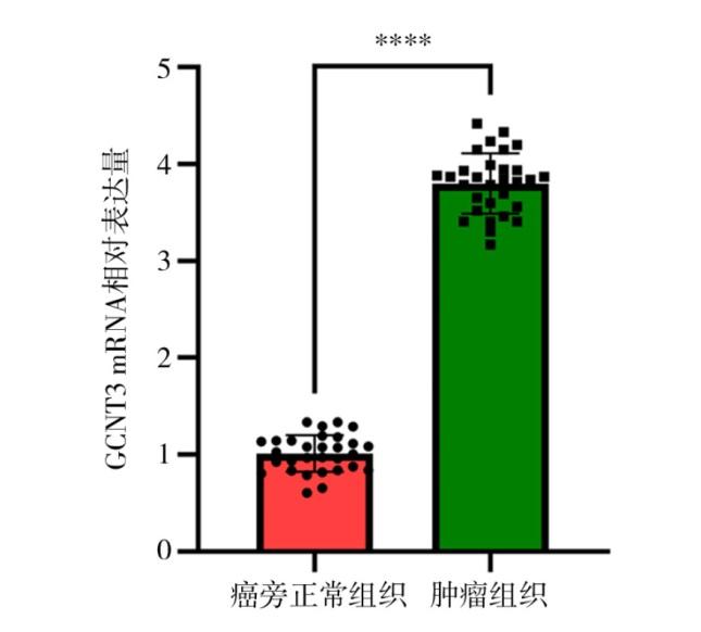
2.1 GCNT3在癌旁正常组织和肺腺癌组织中的表达情况

RT-qPCR法结果显示,癌旁正常组织中GCNT3 mRNA的表达量为1.00±0.23,而肺腺癌组织中GCNT3 mRNA的表达量为3.75±0.28,差异具有统计学意义(t = 41.57,P < 0.000 1)。见图1
图1 GCNT3在癌旁正常组织和肺腺癌组织中的表达情况

Figure 1 Expression of GCNT3 in normal tissue adjacent to cancer and lung adenocarcinoma tissue

注:n = 30;与Normal组比较,****P < 0.000 1。

2.2 下调GCNT3对肺腺癌H1650细胞增殖水平的影响

CCK-8法检测结果显示,Sh NC组在24、48和72 h的增殖率分别为(57.64±3.96)%、(76.26±4.58)%和(92.24±5.12)%;Sh GCNT3组在相应时间点的增殖率分别为(42.32±3.72)%、(62.21±4.62)%和(73.31±4.86)%。组间的增殖率比较差异均有统计学意义(均P < 0.05)。见表1
表1 下调GCNT3对肺腺癌H1650细胞增殖水平的影响

Table 1 The effect of downregulating GCNT3 on the proliferation level of lung adenocarcinoma H1650 cells

组 别 n 细胞增殖率/%
24 h 48 h 72 h
Sh NC组 3 57.64±3.96 76.26±4.58 92.24±5.12
Sh GCNT3组 3 42.32±3.72 62.21±4.62 73.31±4.86
t -4.88 -3.74 -4.64
P 0.008 0.020 0.010

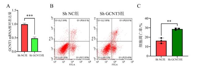
2.3 下调GCNT3对肺腺癌H1650细胞凋亡水平的影响

RT-qPCR法结果显示,与Sh NC组相比,Sh GCNT3组的GCNT3 mRNA表达水平下降,见图2A。进一步流式细胞术结果显示,Sh NC组和Sh GCNT3组的凋亡率分别为(17.46±1.95)%和(30.01±2.63)%。Sh GCNT3组的凋亡率高于Sh NC组,且差异具有统计学意义(t = 6.64,P = 0.003),见图2BC
图2 下调GCNT3对肺腺癌H1650细胞凋亡水平的影响

注:A为RT-qPCR定量分析结果;B为流式细胞仪检测结果,C为CCK-8法定量分析结果;n = 3;与Sh NC组比较,**P < 0.01,***P < 0.001。

Figure 2 The effect of downregulating GCNT3 on the apoptosis level of lung adenocarcinoma H1650 cells

2.4 下调GCNT3对肺腺癌PC3细胞侵袭水平的影响

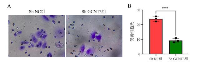
Transwell实验结果显示,2组侵袭细胞数分别为(25.12±2.30)个与(9.26±1.68)个,Sh GCNT3组细胞的侵袭能力低于Sh NC组,差异有统计学意义(t = -9.64,P < 0.001)。见图3
图3 下调GCNT3对肺腺癌H1650细胞侵袭水平的影响

注:A为Transwell实验结果图(结晶紫染色,×400);B为定量分析结果,n = 3,与Sh NC组比较,***P < 0.001。

Figure 3 The effect of downregulating GCNT3 on the invasion level of lung adenocarcinoma H1650 cells

2.5 下调GCNT3对肺腺癌PC3细胞上皮-间质转化表型的影响

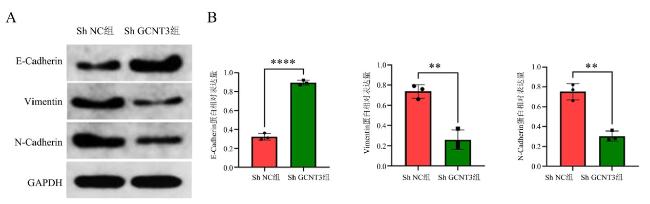
蛋白免疫印迹法结果显示,与NC组(0.33±0.06)比较,Sh GCNT3组E-cadherin蛋白(0.85±0.07)表达水平增加,差异有统计学意义(t = 9.77,P < 0.001);与NC组(0.72±0.06,0.72±0.05)比较,Sh GCNT3组Vimentin、N-cadherin蛋白(0.31±0.05,0.32±0.04)表达水平降低,差异均有统计学意义(t = -9.09,P < 0.001;t = -9.61,P < 0.001)。见图4
图4 下调GCNT3对肺腺癌H1650细胞上皮-间质转化表型的影响

注:A为蛋白免疫印迹法结果;B为定量分析结果,n = 3,与Sh NC组比较,**P < 0.01,****P < 0.000 1。

Figure 4 The effect of downregulating GCNT3 on the EMT phenotype of lung adenocarcinoma H1650 cells

3 讨论

根据近年的全球癌症统计数据,肺癌仍然是导致癌症相关死亡的首要原因,约占全球癌症死亡人数的18%[19-21]。在中国,肺癌也是癌症病死率最高的肿瘤类型,病死率达19%,这一趋势可能与吸烟年轻人群中肺癌发病率的增加密切相关[22-25]。尽管近年来手术、放射治疗、化学治疗及靶向治疗等治疗手段取得了显著进展,肺癌患者的整体预后依然不甚理想,特别是在具有转移、复发及治疗耐药性的患者中[26-30]。肺腺癌是肺癌的一种,属于非小细胞癌。不同于鳞状细胞肺癌,肺腺癌较多见于女性及抽烟者。其起源于支气管黏膜上皮,少数起源于大支气管的黏液腺,其预后优于其他类型的肺癌[31]。探讨肺腺癌放射治疗耐药的机制和潜在分子靶点,对更好地改善患者的生存预后具有重要意义[32]
GCNT3是糖基转移酶家族的重要成员,作为一种黏蛋白型合成酶,GCNT3催化核心2和核心4型O-聚糖的合成,并在蛋白质的O-连接糖基化过程中发挥关键作用[33-35]。多项研究表明,GCNT3在多种癌症中呈现过度表达或突变,与肿瘤的发生和进展密切相关,已被视为潜在的生物标志物和治疗靶点[36-37]。GCNT3在胃癌、结肠癌和卵巢癌等多种恶性肿瘤中起到推动肿瘤进展的重要作用[38-39]。然而,GCNT3在肺腺癌中的具体功能和机制仍有待深入研究。
本研究首先通过RT-qPCR检测了30例肺腺癌及其癌旁正常组织中GCNT3的表达水平,结果显示肺腺癌组织中GCNT3表达高于癌旁正常组织,提示GCNT3可能与肺腺癌的发生密切相关。为进一步探究GCNT3在肺腺癌进展中的作用机制,本研究通过转染GCNT3 ShRNA构建了GCNT3敲低的H1650细胞株,评估了GCNT3的下调对肺腺癌相关恶性生物学行为的影响。结果表明,GCNT3的敲低抑制了H1650细胞的增殖,同时促进了细胞凋亡,这一过程进一步导致了细胞侵袭能力的降低。研究结果表明,GCNT3在调控细胞生长和生存中发挥着关键作用,可能通过影响细胞的生存和迁移机制,协调细胞增殖与凋亡的平衡。因此,GCNT3的表达可能是维持H1650细胞增殖与侵袭能力的重要因素。
EMT是一种细胞通过特定程序由上皮表型转化为间质表型的生物学过程,在肿瘤发展、创伤修复、胚胎发育及器官纤维化过程中具有重要作用[40-42]。EMT通常伴随着上皮细胞黏附分子E-cadherin表达的下调及间质标志物Vimentin的上调,目前被公认是推动肺腺癌转移的重要机制[43-45]。大部分肿瘤细胞中的E-Cadherin的表达是下调的,而N-Cadherin的表达增加[46]。本研究进一步通过蛋白免疫印迹法发现,敲低GCNT3表达水平能够抑制H1650细胞E-cadherin表达的下调、间质标志物Vimentin的上调,而N-Cadherin的表达增加,这表明GCNT3可能通过调控EMT过程促进肺腺癌的侵袭和转移。
综上所述,本研究证实了GCNT3在肺腺癌中的高表达,并通过调控H1650细胞的增殖、侵袭、凋亡及EMT过程,表明GCNT3在肺腺癌的发生、发展中起到至关重要的作用。GCNT3有望作为肺腺癌诊断和治疗的潜在靶点,尤其是在阻止肺腺癌转移方面具有重要的应用前景。
利益冲突声明:本研究未受到企业、公司等第三方资助,不存在潜在利益冲突。
[1]
HAN G, SINJAB A, RAHAL Z, et al. An atlas of epithelial cell states and plasticity in lung adenocarcinoma[J]. Nature, 2024, 627(8004): 656-663. DOI: 10.1038/s41586-024-07113-9.

[2]
LAUGHNEY A M, HU J, CAMPBELL N R, et al. Regenerative lineages and immune-mediated pruning in lung cancer metastasis[J]. Nat Med, 2020, 26(2): 259-269. DOI: 10.1038/s41591-019-0750-6.

PMID

[3]
NICHOLSON A G, TSAO M S, BEASLEY M B, et al. The 2021 WHO classification of lung tumors: impact of advances since 2015[J]. J Thorac Oncol, 2022, 17(3): 362-387. DOI: 10.1016/j.jtho.2021.11.003.

[4]
TRAVIS W D, ASAMURA H, BANKIER A A, et al. The IASLC lung cancer staging project: proposals for coding T categories for subsolid nodules and assessment of tumor size in part-solid tumors in the forthcoming eighth edition of the TNM classification of lung cancer[J]. J Thorac Oncol, 2016, 11(8): 1204-1223. DOI: 10.1016/j.jtho.2016.03.025.

PMID

[5]
SINGH S, BERGSLAND E K, CARD C M, et al. Commonwealth neuroendocrine tumour research collaboration and the North American neuroendocrine tumor society guidelines for the diagnosis and management of patients with lung neuroendocrine tumors: an international collaborative endorsement and update of the 2015 European neuroendocrine tumor society expert consensus guidelines[J]. J Thorac Oncol, 2020, 15(10): 1577-1598. DOI: 10.1016/j.jtho.2020.06.021.

PMID

[6]
FRIEDLAENDER A, PEROL M, BANNA G L, et al. Oncogenic alterations in advanced NSCLC: a molecular super-highway[J]. Biomark Res, 2024, 12(1): 24. DOI: 10.1186/s40364-024-00566-0.

[7]
VLACHOU E, JOHNSON B A 3rd, BARABAN E, et al. Current advances in the management of nonurothelial subtypes of bladder cancer[J]. Am Soc Clin Oncol Educ Book, 2024, 44(3): e438640. DOI: 10.1200/EDBK_438640.

[8]
NASO J R, RODEN A C. Recent developments in the pathology of primary pulmonary salivary gland-type tumours[J]. Histopathology, 2024, 84(1): 102-123. DOI: 10.1111/his.15039.

[9]
WEI X, LI X, HU S, et al. Regulation of ferroptosis in lung adenocarcinoma[J]. Int J Mol Sci, 2023, 24(19): 14614. DOI: 10.3390/ijms241914614.

[10]
WANG Y, FANG X, XIE H, et al. GCNT3 promotes hepatocellular carcinoma progression and EMT by activating the PI3K/AKT pathway[J]. Biochem Genet, 2024. DOI: 10.1007/s10528-024-10830-5.

[11]
ZHANG Y, XIAO P, HU X. LINC00511 enhances LUAD malignancy by upregulating GCNT3 via miR-195-5p[J]. BMC Cancer, 2022, 22(1): 389. DOI: 10.1186/s12885-022-09459-7.

[12]
ZHAO T, ZHAO X, QIAN K, et al. Radiotherapy prognosis-associated gene GCNT3 promotes the proliferation, migration and invasion of lung adenocarcinoma cells[J]. Heliyon, 2022, 8(12): e12100. DOI: 10.1016/j.heliyon.2022.e12100.

[13]
QIU K, WU T, DONG B, et al. GCNT3 regulated MUC13 to promote the development of hepatocellular carcinoma through the GSK3β/β-catenin pathway[J]. Dig Liver Dis, 2024, 56(9): 1572-1581. DOI: 10.1016/j.dld.2024.01.198.

[14]
YAMAMOTO D, SASAKI K, KOSAKA T, et al. Functional analysis of GCNT3 for cell migration and EMT of castration-resistant prostate cancer cells[J]. Glycobiology, 2022, 32(10): 897-908. DOI: 10.1093/glycob/cwac044.

PMID

[15]
LIU J, ZHANG Y, LIU W, et al. miR-BART1-5p targets core 2β-1, 6-acetylglucosaminyltransferase GCNT3 to inhibit cell proliferation and migration in EBV-associated gastric cancer[J]. Virology, 2020, 541: 63-74. DOI: 10.1016/j.virol.2019.12.004.

[16]
FERNÁNDEZ L P, SÁNCHEZ-MARTÍNEZ R, VARGAS T, et al. The role of glycosyltransferase enzyme GCNT3 in colon and ovarian cancer prognosis and chemoresistance[J]. Sci Rep, 2018, 8(1): 8485. DOI: 10.1038/s41598-018-26468-4.

[17]
PASTUSHENKO I, BLANPAIN C. EMT transition states during tumor progression and metastasis[J]. Trends Cell Biol, 2019, 29(3): 212-226. DOI: 10.1016/j.tcb.2018.12.001.

PMID

[18]
HUANG Y, HONG W, WEI X. The molecular mechanisms and therapeutic strategies of EMT in tumor progression and metastasis[J]. J Hematol Oncol, 2022, 15(1): 129. DOI: 10.1186/s13045-022-01347-8.

[19]
HENDRIKS L E L, REMON J, FAIVRE-FINN C, et al. Non-small-cell lung cancer[J]. Nat Rev Dis Primers, 2024, 10(1): 71. DOI: 10.1038/s41572-024-00551-9.

[20]
LOPICCOLO J, GUSEV A, CHRISTIANI D C, et al. Lung cancer in patients who have never smoked-an emerging disease[J]. Nat Rev Clin Oncol, 2024, 21(2): 121-146. DOI: 10.1038/s41571-023-00844-0.

[21]
WANG Z, ZHANG Q, WANG C, et al. Multiple primary lung cancer: updates and perspectives[J]. Int J Cancer, 2024, 155(5): 785-799. DOI: 10.1002/ijc.34994.

[22]
KONDO K K, RAHMAN B, AYERS C K, et al. Lung cancer diagnosis and mortality beyond 15 years since quit in individuals with a 20+ pack-year history: a systematic review[J]. CA Cancer J Clin, 2024, 74(1): 84-114. DOI: 10.3322/caac.21808.

[23]
HE Z, XU Y, RAO Z, et al. The role of α7-nAChR-mediated PI3K/AKT pathway in lung cancer induced by nicotine[J]. Sci Total Environ, 2024, 912: 169604. DOI: 10.1016/j.scitotenv.2023.169604.

[24]
KRATZER T B, BANDI P, FREEDMAN N D, et al. Lung cancer statistics, 2023[J]. Cancer, 2024, 130(8): 1330-1348. DOI: 10.1002/cncr.35128.

[25]
WOLF A M D, OEFFINGER K C, SHIH T Y, et al. Screening for lung cancer: 2023 guideline update from the American Cancer Society[J]. CA Cancer J Clin, 2024, 74(1): 50-81. DOI: 10.3322/caac.21811.

[26]
STRANGE C D, STRANGE T A, ERASMUS L T, et al. Imaging in lung cancer staging[J]. Clin Chest Med, 2024, 45(2): 295-305. DOI: 10.1016/j.ccm.2024.02.004.

PMID

[27]
GUO H, ZHANG J, QIN C, et al. Advances and challenges of first-line immunotherapy for non-small cell lung cancer: a review[J]. Medicine, 2024, 103(3): e36861. DOI: 10.1097/MD.0000000000036861.

[28]
SU P L, CHAKRAVARTHY K, FURUYA N, et al. DLL3-guided therapies in small-cell lung cancer: from antibody-drug conjugate to precision immunotherapy and radioimmunotherapy[J]. Mol Cancer, 2024, 23(1): 97. DOI: 10.1186/s12943-024-02012-z.

[29]
CIARDIELLO F, HIRSCH F R, PIRKER R, et al. The role of anti-EGFR therapies in EGFR-TKI-resistant advanced non-small cell lung cancer[J]. Cancer Treat Rev, 2024, 122: 102664. DOI: 10.1016/j.ctrv.2023.102664.

[30]
BANKS E, YAZIDJOGLOU A, BROWN S, et al. Electronic cigarettes and health outcomes: umbrella and systematic review of the global evidence[J]. Med J Aust, 2023, 218(6): 267-275. DOI: 10.5694/mja2.51890.

PMID

[31]
田甜, 陈港军, 胡创, 等. PIK3R1基因低甲基化在肺腺癌中的临床意义[J]. 暨南大学学报(自然科学与医学版), 2023, 44(4): 358-370. DOI: 10.11778/j.jdxb.20230049.

TIAN T, CHEN G J, HU C, et al. Clinical significance of PIK3R1 gene hypomethylation in lung adenocarcinoma[J]. J Jinan Univ Nat Sci (Med Ed), 2023, 44(4): 358-370. DOI: 10.11778/j.jdxb.20230049.

[32]
钱晶, 何泽来, 田校源, 等. 68例EGFR突变型肺腺癌脑转移患者早期颅脑放疗对比延迟放疗疗效分析[J]. 中华全科医学, 2021, 19(12): 1995-1998, 2027. DOI: 10.16766/j.cnki.issn.1674-4152.002219.

QIAN J, HE Z L, TIAN X Y, et al. Efficacy analysis of early radiotherapy versus delayed radiotherapy in 68 cases of EGFR-mutant lung adenocarcinoma with brain metastases[J]. Chin J Gen Pract, 2021, 19(12): 1995-1998, 2027. DOI: 10.16766/j.cnki.issn.1674-4152.002219.

[33]
GUPTA R, LEON F, THOMPSON C M, et al. Global analysis of human glycosyltransferases reveals novel targets for pancreatic cancer pathogenesis[J]. Br J Cancer, 2020, 122(11): 1661-1672. DOI: 10.1038/s41416-020-0772-3.

[34]
RAO C V, JANAKIRAM N B, MOHAMMED A. Molecular pathways: mucins and drug delivery in cancer[J]. Clin Cancer Res, 2017, 23(6): 1373-1378. DOI: 10.1158/1078-0432.CCR-16-0862.

PMID

[35]
XI X, WANG J, QIN Y, et al. Glycosylated modification of MUC1 maybe a new target to promote drug sensitivity and efficacy for breast cancer chemotherapy[J]. Cell Death Dis, 2022, 13(8): 708. DOI: 10.1038/s41419-022-05110-2.

[36]
AGOSTINI A, GUERRIERO I, PIRO G, et al. Talniflumate abrogates mucin immune suppressive barrier improving efficacy of gemcitabine and nab-paclitaxel treatment in pancreatic cancer[J]. J Transl Med, 2023, 21(1): 843. DOI: 10.1186/s12967-023-04733-z.

[37]
杨永泉, 田永靖, 庞健, 等. 肿瘤相关巨噬细胞通过诱导GCNT3上调促进胃癌细胞上皮-间质转化[J]. 中国免疫学杂志, 2023, 39(3): 483-488. DOI: 10.3969/j.issn.1000-484X.2023.03.006.

YANG Y Q, TIAN Y J, PANG J, et al. Tumor-associated macrophages promote epithelial-mesenchymal transition of gastric cancer cells by inducing GCNT3 up-regulation[J]. Chin J Immunol, 2023, 39(3): 483-488. DOI: 10.3969/j.issn.1000-484X.2023.03.006.

[38]
刘陕西, 叶钧, 尚杨杨, 等. GCNT3在结直肠癌中的表达及临床意义[J]. 第三军医大学学报, 2019, 41(10): 913-917. DOI: 10.16016/j.1000-5404.201812043.

LIU S X, YE J, SHANG Y Y, et al. Expression and clinical significance of GCNT3 in colorectal cancer[J]. J Third Mil Med Univ, 2019, 41(10): 913-917. DOI: 10.16016/j.1000-5404.201812043.

[39]
孙晓燕, 刘畅, 张华, 等. GCNT3表达在非小细胞肺癌中的临床意义[J]. 中国肿瘤临床, 2019, 46(3): 111-116. DOI: 10.3969/j.issn.1000-8179.2019.03.377.

SUN X Y, LIU C, ZHANG H, et al. Clinical significance of GCNT3 expression in non-small cell lung cancer[J]. Chin J Clin Oncol, 2019, 46(3): 111-116. DOI: 10.3969/j.issn.1000-8179.2019.03.377.

[40]
SAITOH M. Transcriptional regulation of EMT transcription factors in cancer[J]. Semin Cancer Biol, 2023, 97: 21-29. DOI: 10.1016/j.semcancer.2023.10.001.

PMID

[41]
AKHMETKALIYEV A, ALIBRAHIM N, SHAFIEE D, et al. EMT/MET plasticity in cancer and Go-or-Grow decisions in quiescence: the two sides of the same coin[J]. Mol Cancer, 2023, 22(1): 90. DOI: 10.1186/s12943-023-01793-z.

[42]
AKRIDA I, PAPADAKI H. Adipokines and epithelial-mesenchymal transition (EMT) in cancer[J]. Mol Cell Biochem, 2023, 478(11): 2419-2433. DOI: 10.1007/s11010-023-04670-x.

[43]
SALEEM H M, RAMAIAH P, GUPTA J, et al. Nanotechnology-empowered lung cancer therapy: from EMT role in cancer metastasis to application of nanoengineered structures for modulating growth and metastasis[J]. Environ Res, 2023, 232: 115942. DOI: 10.1016/j.envres.2023.115942.

[44]
XIE S, WU Z, QI Y, et al. The metastasizing mechanisms of lung cancer: Recent advances and therapeutic challenges[J]. Biomed Pharmacother, 2021, 138: 111450. DOI: 10.1016/j.biopha.2021.111450.

PMID

[45]
MAK M P, TONG P, DIAO L, et al. A patient-derived, pan-cancer EMT signature identifies global molecular alterations and immune target enrichment following epithelial-to-mesenchymal transition[J]. Clin Cancer Res, 2016, 22(3): 609-620. DOI: 10.1158/1078-0432.CCR-15-0876.

PMID

[46]
陈文灿, 周倜. N-钙黏蛋白功能研究进展[J]. 中山大学学报(医学科学版), 2024, 45(6): 866-875. DOI: 10.13471/j.cnki.j.sun.yat-sen.univ(med.sci).20241030.002.

CHEN W C, ZHOU T. Research progress in N-cadherin function[J]. J Sun Yat-sen Univ (Med Sci), 2024, 45(6): 866-875. DOI: 10.13471/j.cnki.j.sun.yat-sen.univ(med.sci).20241030.002.

Outlines

/